
The abnormal blood vessels associated with diabetic retinopathy stimulate the growth of scar tissue, which can pull the retina away from the back of the eye. Unless your retina is damaged, your vision will likely return to its previous clarity. Ultimately, the retinologist used a combination of procedures to repair the damage including: 1) scleral buckling 2) pneumatic. The blood often clears from the eye within a few weeks or months. The retinologist stated that he would place a scleral buckle in the eye to repair the detachment, that the patient would have to lie in a prone position (stomach) for a minimum of three days, and could only get up to go to the bathroom and eat. If we do not do this now, you will be blind. The patient then asked when the surgery would be performed the retinologist stated tomorrow morning at 7:00.

The retinologist examined both eyes and stated that you have a retinal detachment with multiple tears in the right eye that will require surgery to repair. On Monday morning, a visit to the retinologist was scheduled for the following Thursday afternoon. The NEI says, Symptoms include a sudden or gradual increase in either the number of floaters, which are little cobwebs or specks that float about in your. This blackened area began to slowly extend to cover more of the eye. The following day, the patient noticed that the medial corner of the eye near the nose developed a blackened area. The following day, the patient began to notice a "shower" of "floaters" in the right eye though no action was taken by the patient. A detached retina needs treatment as soon as possible. Sudden changes, including eye floaters and flashes and darkening side vision, are signs this may be happening. The retina, the layer of tissue in the back of the eye, pulls away from tissues supporting it. The examination was completed and new glasses were ordered due to a change in vision. Retinal detachment, or a detached retina, is a serious eye condition. Follow-up visits were scheduled and the hemorrhage repaired itself without intervention.ĭuring the last week of August of that same year, the patient scheduled an exam with the ophthalmologist to determine the need for new glasses. Often, a retinal tear precedes full-on detachment, and catching it at this stage is the better scenario, Dr. A return visit was scheduled and upon examination, the patient was told the same information as stated above, except for the fact that there was now a posterior vitreous hemorrhage (PVH) in the left eye. However, the retinologist stated that the patient should return for a follow-up visit at six months or to return earlier should significant changes be noticed.Īpproximately four months later, this patient noticed that "floaters" began to appear in the right eye. The patient continued to observe the "floaters" which did vary in number and size over the ensuing four months. Upon completion of the evaluation, the retinologist told the patient that these materials were termed "floaters" and were insignificant, required no treatment, and would resolve over time by either sinking to the bottom of the vitreous humor, or the brain would learn to ignore them. Luckily, the retinologist was able to work the patient in that same morning. Immediately upon the mention of these materials floating around in the eye, the patient was informed that the ophthalmologist would not see the patient and would be referring him to a retinologist. On the day of the appointment with the ophthalmologist, his medical assistant began to take a patient history. The materials noted varied from time to time but appeared as discrete black balls of varying sizes, spider web-like materials, and irregularly-shaped materials that were not discrete. This can cause irregular blood flow which may lead to clotting.A 71-year-old white male began to observe materials floating around in his left eye in early January and called to make an appointment with his ophthalmologist.
.jpg "webmd detached retina symptoms")
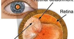
When an artery hardens, it can press against a vein and narrow the opening. In the retina, arteries and veins cross over each other. Retinal vein occlusion usually occurs due to a blood clot that blocks the vein. The blockage of the vein means that blood cannot drain out of the retina. Retinal vein occlusion occurs when a vein draining blood from the eye gets blocked. The three main variations of retinal detachment include: Brief, bright flashes of light These flashes may be most. These are actually red blood cells because all retinal tears bleed a little when they occur. Most worrisome is a shower of black dots. Retinal detachment occurs when the retina becomes separated from the underlying supportive tissues, which prevents it from functioning correctly. Outlook Retinal detachment occurs when the retina, a light-sensitive membrane, separates from the back of the eye. Symptoms of a detached retina may include: The sudden appearance of 'floaters' (dark, semi-transparent, floating shapes) in the field of vision.
 0 kommentar(er)
